
Digital Diagnosis and Planning
Dental Radiology / CBCT
Radiological imaging in dentistry is used to assess tooth and bone structures that cannot be seen directly at a clinical examination. The type of imaging should be chosen according to the clinical question; not carrying out unnecessary imaging is essential.
Medical review: Dr. Taylan Akça. Last updated: Aug. 29, 2026.
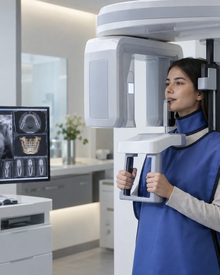
Panoramic X-ray
This is a two-dimensional image that helps to assess all of the jaws and teeth in general. It is useful for impacted teeth, the overall bone structure and assessing many teeth together.
CBCT
Cone beam computed tomography (CBCT) provides three-dimensional information in selected situations. It may be used in implant planning, the relationship between an impacted tooth and a nerve, some surgical lesions and assessing anatomical structures.
Frequently asked questions
Is CBCT needed for every implant?
The need is assessed according to the case and the region. If three-dimensional anatomical information affects the treatment decision, a CBCT may be requested.
Is radiation dangerous?
For all ionising radiation procedures, justification and the principle of the lowest appropriate dose possible are important. Imaging is chosen according to the clinical requirement.
Is an X-ray taken during pregnancy?
An urgent need and a risk–benefit assessment should be made by the clinician; pregnancy or suspected pregnancy must be reported.
Treatment duration
The imaging procedure is short; which imaging is needed is determined by the clinical requirement.
When it may not be suitable
For all ionising radiation procedures, justification and the principle of the lowest appropriate dose possible are important. Pregnancy or suspected pregnancy must be reported.